 by
by An imaging sensor inspired by the visual system of butterflies has been developed by a team of researchers. The sensor is capable of detecting ultraviolet (UV) light, a range that is inaccessible to human eyes. The design of the sensor uses stacked photodiodes and perovskite nanocrystals (PNCs) to image different wavelengths in the UV range. By utilizing the spectral signatures of biomedical markers such as amino acids, the imaging technology can differentiate between cancer cells and normal cells with 99% accuracy.
The research, led by professors Viktor Gruev and Shuming Nie from the University of Illinois Urbana-Champaign, was published in the journal Science Advances.
UV light is electromagnetic radiation with shorter wavelengths than visible light and longer than x-rays. The ability to capture UV information and differentiate between sub-regions such as UVA, UVB, and UVC is challenging for humans due to our inability to see UV light. However, butterflies can perceive small variations in the UV spectrum, similar to how humans perceive shades of blue and green. The researchers were particularly intrigued by how butterflies can capture UV light, as it tends to be absorbed easily.
Butterflies have compound eyes with six or more photoreceptor classes that possess distinct spectral sensitivities. The Papilio xuthus butterfly, for example, has photoreceptors for blue, green, red, violet, ultraviolet, and broadband wavelengths. Additionally, butterflies have fluorescent pigments that convert UV light into visible light, enhancing their ability to perceive a broader range of colors and details in their environment.
To replicate the UV sensing mechanism of the Papilio xuthus butterfly, the UIUC team combined a thin layer of PNCs with a tiered array of silicon photodiodes. PNCs are semiconductor nanocrystals with properties akin to quantum dots, allowing them to absorb and emit light of different wavelengths based on their composition. The PNC layer in the imaging sensor absorbs UV photons and re-emits them as visible green light, which is then detected by the tiered silicon photodiodes. Signal processing enables the mapping and identification of UV signatures.
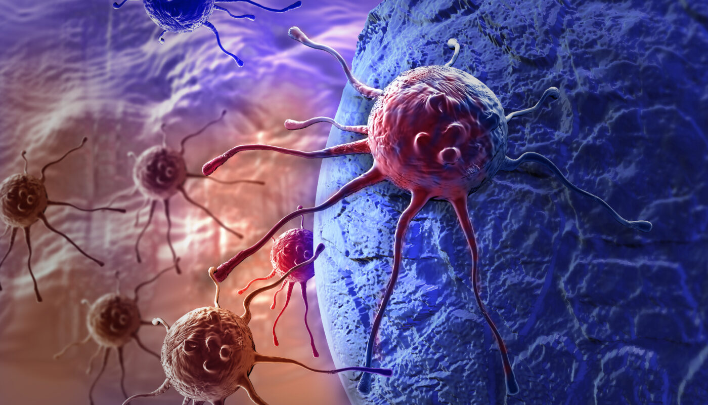
In terms of applications, the ability to detect UV light with high sensitivity and distinguish small differences in wavelength opens up possibilities in healthcare and other fields. Cancerous tissues have higher concentrations of certain biomedical markers, such as amino acids, proteins, and enzymes, compared to healthy tissues. When excited with UV light, these markers fluoresce in the UV and visible spectrum. The imaging device developed by the research team can differentiate between cancer and healthy cells with 99% confidence based on their fluorescence in the UV spectrum.
The researchers envision using the sensor during surgeries to aid in decision-making processes, particularly in determining the extent of tissue removal to ensure clear margins when removing cancerous tumors.
Beyond healthcare, the butterfly-inspired imaging sensor can also be used in various other applications such as biology and underwater exploration. By detecting UV light, researchers can gain insights into the behavior of species that can perceive and use UV light, including their hunting and mating habits. Additionally, studying the impact of UV light underwater can provide a greater understanding of the underwater environment and the ways in which animals utilize UV light, despite its partial absorption by water.
*Note:
1. Source: Coherent Market Insights, Public sources, Desk research
2. We have leveraged AI tools to mine information and compile it